

Case
MTA Plug Apexification
This case report highlights the non-surgical management of a non-vital tooth with an open apex associated with a peri-apical lesion using MTA apical plug technique.
MTA plug in the apical portion of the root promotes apical repair and provides an apical stop. MTA has an excellent sealing ability and sets even in the presence of moisture.
Apical gauging is essential to determine the narrowest diameter of the prepared canal and the apical constriction. A number of chemical mediators related to the pathogenesis of periapical lesions stimulate resorption of the root as well as the bone, so destroying the constriction. This is known known as apical inflammatory root resorption.
Destruction of apical constriction and can lead to extrusion of the root canal filling through the apex and inability to create a good apical seal which is essential to a successful outcome.
Case presentation:
The following case presents a young lady requesting cosmetic treatment of upper central incisors. She suffered trauma to the upper incisors aged 8 and the teeth had discoloured. On examination a draining sinus and large periapical areas were associated with upper right central incisor (UR1) and upper left central incisor (UL1).
Diagnosis:
Upper right central incisor (UR1) and upper left central incisor (UL1) non vital with associated chronic apical abscess at UR1 and chronic periapical periodontitis at UL1.
Treatment:
Root canal treatment was initiated, and apical gauging found the apex of UR1 and UL1 to be wider than 0.5 mm (iso size 50 file) The normal size of the apical constriction is between 0.15 mm and 0.20mm.
A diagnosis of apical inflammatory resorption was made and due to concerns of overfilling and the ability of gutta percha to produce a long-term apical seal, an MTA plug was placed in the apical 4 mm of UR1 and UL1.
On setting the canals were then back filled with heated gutta percha and AH Plus sealer.
At 4 week review the sinus had completely healed and the UL1 and UR1.
At 12 month review the radiograph shows bone healing.
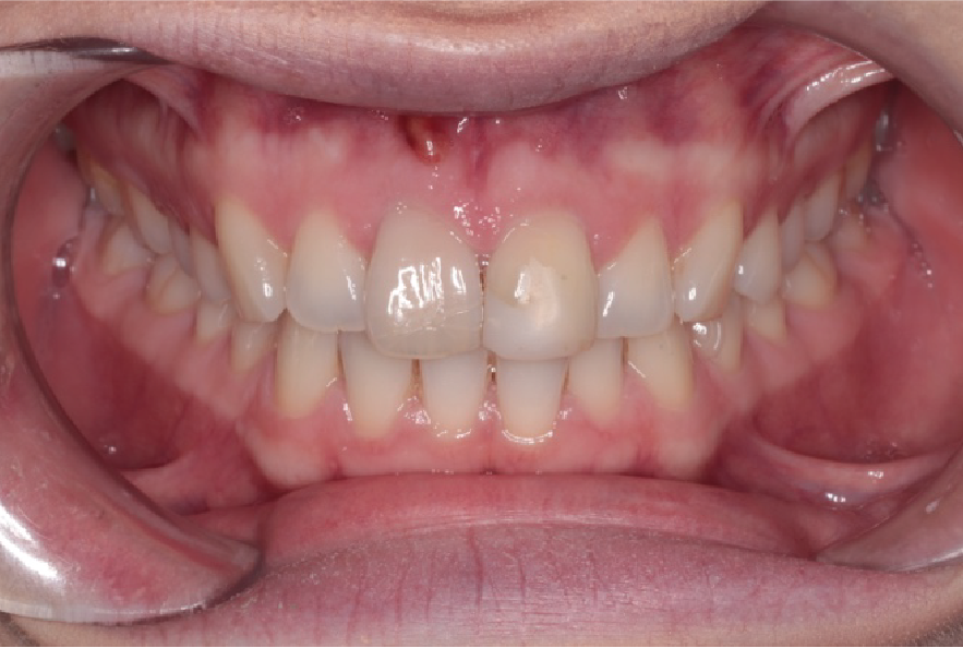
Pre-operative photo, showing sinus apical to UR1.

Right lateral showing sinus apical to UR1.